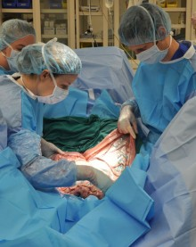
CALEC Surgery: A Breakthrough in Eye Injury Treatment
CALEC surgery, a groundbreaking procedure recently implemented at Mass Eye and Ear, offers newfound hope for individuals suffering from severe corneal damage. This innovative surgery utilizes cultivated autologous limbal epithelial cells to regenerate the cornea, a critical component of eye health. In a clinical trial, stem cell therapy successfully restored the cornea’s surface in patients with blinding injuries, showcasing a remarkable success rate of over 90%. The procedure not only highlights advancements in corneal damage treatment but also opens the door for enhanced eye injury recovery through cutting-edge techniques. As CALEC surgery continues to evolve, it paves the way for transformative approaches to preserve and restore vision, significantly benefiting those who had previously lost hope.
The cultivation of limbal epithelial cells represents a major leap in ocular medicine, reflecting a commitment to innovative approaches in healing eye injuries. Known as CALEC surgery, this technique harnesses the power of stem cell therapy to restore corneal integrity and functionality. With its roots in advanced clinical trials, this procedure not only targets chronic corneal damage but presents a viable alternative to traditional transplantation methods. The Mass Eye and Ear trial highlights the potential for this approach to change the face of corneal restoration, offering new possibilities for those affected by severe eye conditions. As researchers continue to explore this exciting field, the prospects for comprehensive eye care are brighter than ever.
Understanding CALEC Surgery: A Breakthrough in Eye Treatment
Cultivated autologous limbal epithelial cell (CALEC) surgery represents a revolutionary advancement in the treatment of severe corneal damage. This innovative technique involves harvesting limbal epithelial cells from a healthy eye and cultivating them to create a tissue graft. The process, although intricate, restores the surface of the damaged cornea, which can lead to substantial improvements in vision and quality of life for patients suffering from corneal ailments. With over 90 percent success rate reported in the recent trials at Mass Eye and Ear, Ula Jurkunas and her team have not only established the feasibility of CALEC surgery but have also showcased its potential in transforming how we address previously untreatable cases of eye injury and damage.
The limbus contains valuable stem cells that are crucial for maintaining the health and transparency of the cornea. When these stem cells are depleted due to injuries like chemical burns or infections, conventional treatments often fail, leaving patients with chronic pain and vision loss. CALEC surgery offers a beacon of hope, allowing for the regeneration of these essential cells, thereby re-establishing the corneal surface’s integrity. Trials involving CALEC have highlighted not just its safety but also its potential to avert future complications associated with traditional treatments such as corneal transplants.
As a pioneering solution in the field of corneal damage treatment, CALEC surgery is set against the backdrop of a growing need for more effective therapies. Traditional methods, often reliant on corneal transplants, come with limitations, particularly in cases where patients may have damage to both eyes. The innovative approach demonstrated in the Mass Eye and Ear trials opens avenues for future research and potential standardization of the procedure within ophthalmology. Ula Jurkunas’s insight into the application of stem cell therapy in corneal restoration exemplifies how modern techniques in regenerative medicine are reshaping the management of severe ocular conditions.
How Stem Cell Therapy Revolutionizes Corneal Damage Treatment
The integration of stem cell therapy into corneal damage treatment marks a significant turning point in how we understand and address eye injuries. At the heart of this revolution lies the concept of harnessing the body’s own regenerative capabilities. By isolating limbal epithelial cells from a healthy eye, researchers can cultivate them in a controlled environment, essentially creating a personalized tissue graft that is then transplanted into the injured eye. This innovative method demonstrates not only the effectiveness of stem cell therapy in treating corneal damage but also its safety profile, as evidenced by the outcomes of the Mass Eye and Ear trials, which report high success rates over extended follow-up periods.
Moreover, the empowering aspect of stem cell therapy lies in its potential to alleviate chronic conditions stemming from corneal injuries. Patients who once faced severe visual impairment due to conditions like limbal stem cell deficiency can now aspire to regain their sight without undergoing invasive traditional surgeries. This paradigm shift not only reduces the risks associated with corneal transplants but also enhances the quality of life for many, highlighting the transformative impact that stem cell therapy has in modern ophthalmology.
Furthermore, the application of stem cell therapy extends beyond just individual treatments; it prompts a re-evaluation of the entire corneal repair strategy. While conventional methods often involve a one-size-fits-all approach, the personalized nature of stem cell treatments like CALEC surgery allows for tailored interventions that address the unique needs of each patient. This shift towards individualized therapies opens doors for more comprehensive care pathways in ophthalmology, potentially facilitating quicker recovery times and minimizing the risk of complications. As research continues to evolve, the future of corneal damage treatment is increasingly centered on harnessing regenerative medicine to restore vision and improve patient outcomes.
The Process Behind CALEC Surgery: From Lab to Clinic
The journey of CALEC surgery from concept to clinical application involves meticulous research, innovation, and collaboration among leading experts. This surgical procedure begins with the extraction of a small tissue sample from the healthy eye, where limbal epithelial cells reside. These cells are then cultured in a specialized environment where they proliferate, creating a graft that is rich in the necessary stem cells. This advanced manufacturing process, taking two to three weeks, ensures that the graft is of high quality and suitable for transplantation back into the affected eye. The meticulous nature of this process not only highlights the scientific rigor behind CALEC surgery but also underscores its potential to provide sustainable solutions for patients experiencing severe corneal impairments.
Another crucial aspect of CALEC surgery’s success is the collaborative environment at Mass Eye and Ear, where surgeons, researchers, and manufacturing experts come together. The synergy between the clinical and research teams, including the partnerships with well-respected institutions such as Dana-Farber and Boston Children’s Hospital, has been integral to the development and application of this innovative therapy. The trials, which began with stringent quality oversight and safety evaluations, have now positioned CALEC as a leader in the field of surgery for corneal repair, paving the way for future advancements in stem cell therapies and their application in other areas of ophthalmology.
Transitioning from laboratory successes to real-world applications, CALEC surgery also represents a unique opportunity to further study the regenerative properties of limbal epithelial cells in treating various ocular conditions. While the current trial has shown promising results, the continued evolution of this technique will rely on expanded studies that encompass larger, diverse patient populations. By advancing toward longer follow-up periods and randomized-control designs, researchers aspire to gather comprehensive data that can fortify the case for CALEC as a mainstream treatment option. This progression is vital for achieving FDA approval, which could subsequently broaden accessibility to such innovative therapies and empower countless patients suffering from corneal damage.
Success Rates and Outcomes of CALEC Surgery
The remarkable outcomes of CALEC surgery, particularly delineated in the recent clinical trials, accentuate its effectiveness in restoring corneal surfaces in patients with severe eye damage. As indicated by findings from ongoing assessments, approximately 50 percent of participants experienced full restoration of the cornea within three months post-surgery, a number that bolstered to nearly 79 percent at the one-year mark. These statistics reflect not only the procedure’s high efficacy but also its potential to significantly enhance the overall quality of life for individuals who had previously faced debilitating vision loss due to corneal damage. Such success rates underscore the necessity for further exploration and potentially wider implementation of CALEC in clinical practices across the nation.
Moreover, the incremental increases in success rates showcase the therapy’s capability to adapt and respond to individual recovery patterns. With observed improvements in visual acuity among all participants, the CALEC procedure indicates a strong safety profile, further solidifying its place in the realm of advanced ocular therapies. It is crucial for both medical professionals and patients to recognize the transformative potential of CALEC surgery as a beacon of hope for restorative eye treatment. Given the compelling evidence emerging from the clinical trials, the future looks promising for this innovative therapy, inspiring continued research and shedding light on new interventions that aim to address corneal defects.
As the results from the Mass Eye and Ear trial delineate, the journey of recovery after CALEC surgery is multifaceted, involving not only the restoration of the corneal surface but also the potential for improved vision. Importantly, the reported complications were minimal, primarily involving manageable conditions that resolved swiftly following treatment. This speaks volumes about the robustness of CALEC surgery and its safety in comparison to conventional therapeutic options. With the knowledge gained from these preliminary findings, there is an escalating interest among eye care specialists and patients alike regarding the future of CALEC surgery in advancing standard care practices for corneal injuries, thereby promising a new chapter in regenerative ophthalmology.
The Future of Limbal Stem Cell Therapy
The future of limbal stem cell therapy, particularly through innovations like CALEC surgery, is poised for significant advancements as ongoing research and trials unfold. The aim is to broaden the application of stem cell-based techniques by exploring an allogeneic manufacturing process that involves sourcing limbal stem cells from cadaveric donor eyes. This evolutionary step could help extend the reach of treatments to patients suffering from bilateral corneal damage, thereby increasing the overall impact of this therapy in the field of ophthalmology. The enthusiasm surrounding these developments is supported by the impressive outcomes recorded during the initial trials, which could serve as a blueprint for further exploration in similar regenerative fields across medicine.
There is also an urgent need for continued vigilance in research practices to ensure that the transition from experimental procedures to approved therapies remains consistent and safe. As researchers target larger populations and more robust trial designs, the hope is to secure FDA approval, ultimately enabling a broader dissemination of this groundbreaking treatment across the United States. The implications of successful FDA endorsement for CALEC surgery could transform the therapeutic landscape for corneal injuries and other ocular manifestations, propelling regenerative medicine to the forefront of eye care.
One of the most significant aspects of future limbal stem cell therapy involves the commitment to rigorous scientific inquiry while maintaining patient safety and quality care as paramount. As collaborative efforts among institutions like Mass Eye and Ear and partnerships with organizations like the National Eye Institute continue, the stream of innovation and success in the field can be expected to flourish. The vision of making living cell-based therapies widely available to patients on a national scale is no longer just a dream; it is an achievable reality that reflects the ongoing dedication to improving treatment outcomes in ophthalmology.
Frequently Asked Questions
What is CALEC surgery and how does it help in treating corneal damage?
CALEC surgery, which stands for Cultivated Autologous Limbal Epithelial Cells surgery, involves harvesting stem cells from the limbus of a healthy eye and transplanting them into a damaged eye. This innovative procedure helps restore the cornea’s surface, offering new hope for patients with severe corneal damage that was previously deemed untreatable.
What role does stem cell therapy play in CALEC surgery?
Stem cell therapy is a critical component of CALEC surgery. It allows for the regeneration of limbal epithelial cells, which are essential for maintaining the cornea’s clarity and health. By using stem cells harvested from a healthy eye, CALEC therapy aims to successfully repair the damaged corneal surface and improve visual outcomes for patients.
Can CALEC surgery be used for patients with eye injuries?
Yes, CALEC surgery is specifically designed for patients suffering from corneal damage due to eye injuries, chemical burns, infections, or other traumas. This surgery utilizes cultivated stem cells to regenerate the corneal surface, providing an innovative treatment option for those with significant eye injuries.
What were the results of the Mass Eye and Ear trial involving CALEC surgery?
The clinical trial at Mass Eye and Ear demonstrated that CALEC surgery is safe and effective, with a success rate of over 90% in restoring the cornea’s surface among participants over an 18-month period. The study involved 14 patients and showcased significant improvements in visual acuity following the procedure.
How do limbal epithelial cells contribute to corneal health in CALEC surgery?
Limbal epithelial cells are crucial for the health of the cornea as they maintain its smooth surface. In CALEC surgery, these stem cells are cultivated from a healthy eye and transplanted to the damaged cornea to restore its integrity and function, thereby improving patients’ vision.
Are there any risks associated with CALEC surgery?
While CALEC surgery has shown a strong safety profile, it is still considered experimental and involves some risks. During the trial, there was one reported bacterial infection in a participant, but no serious adverse events were linked to the procedure itself. Patients need to be thoroughly evaluated before considering this treatment.
What is the future of CALEC surgery and its availability?
The future of CALEC surgery looks promising, with ongoing studies aimed at increasing patient access and expanding the treatment’s applicability, including a potential allogeneic approach. However, as of now, CALEC surgery is not widely available, as further trials and regulatory approvals are necessary.
Who conducted the research on CALEC surgery and where was it tested?
The research on CALEC surgery was conducted by Ula Jurkunas and her team at Mass Eye and Ear, in collaboration with Dana-Farber Cancer Institute. The trial represents a significant advancement in the field of ophthalmology, being the first human study of its kind funded by the National Eye Institute.
| Key Points |
|---|
| First CALEC surgery performed at Mass Eye and Ear by Ula Jurkunas. |
| Procedure harvested stem cells from a healthy eye and transplanted them into a damaged eye, restoring corneal surfaces in 14 patients. |
| CALEC is over 90% effective at restoring the cornea’s surface, a significant advancement for those with previously untreatable corneal damage. |
| Study monitored patients for 18 months, showing gradual improvement in corneal health and vision. |
| No serious adverse events related to the treatment; minor issues resolved quickly. |
| Future studies aim to broaden the applicability of CALEC and eventually seek FDA approval. |
Summary
CALEC surgery represents a groundbreaking advancement in the field of ophthalmology, offering new hope for individuals suffering from corneal damage that was once deemed untreatable. This innovative procedure utilizes stem cells to restore the cornea’s surface effectively, achieving over 90% success rates in clinical trials. As research continues, the future of CALEC surgery holds promise for broader accessibility and improved treatment outcomes for patients with eye injuries.
